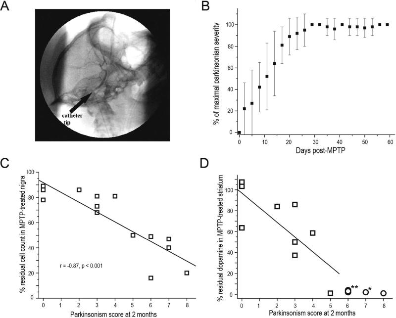
Figure 5.
PET correlates with nigrostriatal damage and parkinsonism following unilateral intracarotid MPTP in non-human primates. A) Digital subtraction lateral projection cerebral angiography in a non-human primate demonstrating the location of the unilateral intra-carotid catheter tip just prior to injection of MPTP. B) Plot demonstrating the behavioral response in different animals given varying doses of MPTP (y axis = degree of parkinsonism) over 2 months (x axis = time) following unilateral intra-carotid MPTP administration. Error bars represent standard deviations (N=15). C) Parkinsonism score at 2 months versus percent of residual dopaminergic cell counts and D) dopamine content in the striatum (n=14). Data from monkeys with residual nigral cell counts of less than 50% are depicted by circles (n=5). Squares represent data from monkeys with residual nigral cell counts of 50% or more (n=9). Figure and legend adapted from Tabbal et al., 2012.26

