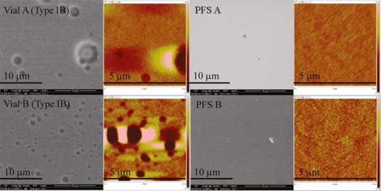
Fig. 8.

SEM (grayscale) and AFM images (color) of vials and PFS after exposure to pH 11 glutaric acid at 40°C/75% RH for 3 months. The depth scale for AFM images is from −20 to 20 nm for Vial A Type IB, −10 to 10 nm for Vial B Type IB, and −5 to 5 nm for PFS A and B
