
Fig. 9.
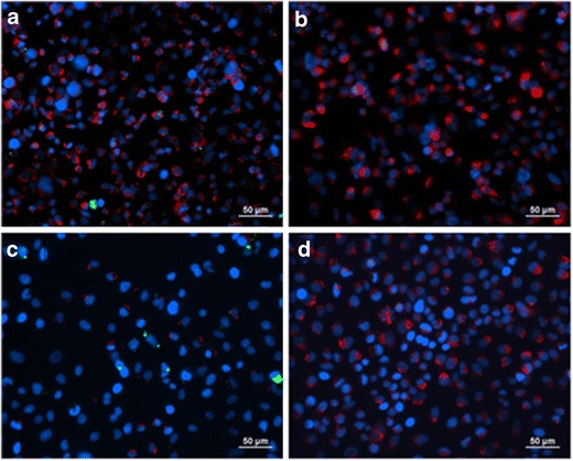
Merged fluorescent micrographs of A549 lung adenocarcinoma cells exposed to co-SD 50PTX:50DPPC/DPPG formulated particles loaded with 1 mol% NBD-PC (fluorophore) for 6 h (a and b, top row) and 24 h (c and d, bottom row) at 37°C: a and c show the micrographs of cells that were exposed to particles, and b and d are control cells with no particle exposure. The nucleus (blue) and cytoplasm (red) were fluorescently labeled after particle exposure and washing