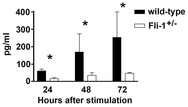
Fig. 3. Decreased IL-6 production in T cells isolated from Fli-1+/− MRL/lpr mice compared to wild-type littermates.

T cells were negatively isolated from spleens of Fli-1+/− (n=6) or wild-type littermates (n=6) at the age of 16 weeks using the T cell negative Dynabead isolation kit. The T cells were cultured in 24-well plates at a concentration of 1×10⁶/ml cells. The supernatants were collected 1 to 3 days after stimulation with CD3/CD28 activator microbeads and ELISA was used to determine the IL-6 concentrations. The data presented are the mean ± SD. * p<0.05.
