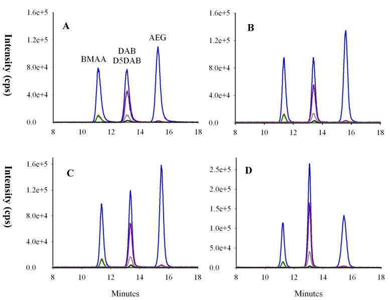
Figure 5.
Chromatograms of BMAA, D5DAB and AEG spiked at 50 ng/mL into (A) standards; (B) cyanobacteria; (C) oyster and (D) mussel matrices after extraction of free analytes. Colored lines represent mass spectral transitions at m/z 119 to m/z 102 (blue), 88 (red), 76 (green), 101 (purple), 74 (grey) and m/z 124 to m/z 47 (dark).

