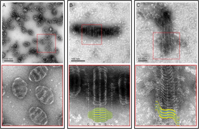
Figure 2.
Representative structures of the 6-His-MVP mutant. Electron micrographs of uranyl acetate stained supernatants of lysates from infected Sf9 cells. (A) Wild-type MVP vaults with a close-up view from its red inset. (B) Staggered rolls of MVP chains. Close-up view of the rolls aligned with a vault particle from the crystal structure.10 The vault cap (C), shoulder (S), and waist (W) regions are indicated by white dashed lines. (C) Long sheet of an unraveled MVP roll. Close-up view of the sheet superimposed with several individual MVP chains from the crystal structure.

