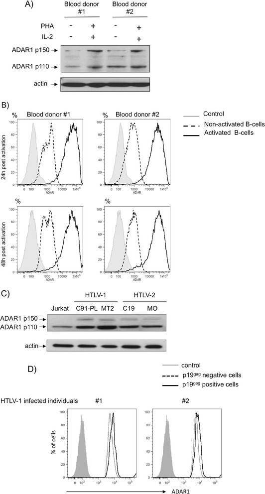
Figure 1.

ADAR1 is expressed in activated lymphocytes as well as in HTLV-1 and HTLV-2 chronically infected T-cell lines and primary T-lymphocytes from HTLV-1 infected individuals. (A): Whole cell extracts (60 μg) obtained from PBLs activated or not with PHA/IL-2 for 72 hours. (B): B-cells were purified from the PBMCs of two blood donors and left unactivated (dot line) or treated with Pansorbin (solid line). ADAR1 expression was measured 24 h and 48 h later (FACS Canto II, BD Biosciences). Isotype control is represented with a grey histogram (C): Uninfected- (Jurkat), HTLV-1-infected (C91-PL, MT2) and HTLV-2-infected (C19, MO) cells were analyzed by western-blot analyses using anti-ADAR1 or anti-actin antibody. (D): CD4+ T-cells from two HTLV-1 positive individuals were sorted, stimulated with PHA and cultured in the presence of IL-2. Cells were stained with anti ADAR1 and anti HTLV p19gag antibodies followed by APC anti-rabbit and FITC anti-mouse antibodies. CD4+ cells from p19gag negative population (dot line) or from p19gag positive population (solid line) were analyzed for ADAR1 expression. (B, D): Cells were analyzed on a FACS-Canto II (BDSciences) collecting 100 000 events. Results analyzed using FlowJo software.
