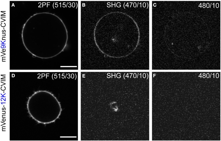
Figure 3.
Second harmonic imaging of mVe9Knus-CVIM. (A–C) Images showing two-photon excited fluorescence (2PF), SHG, and off-band signals from a cell expressing mVe9Knus-CVIM, respectively. (D–F) Same for a cell expressing mVenus-12K-CVIM. Fundamental wavelength was 940 nm. (B,C,E,F) Used the same brightness/contrast settings to allow direct compa-risons. Actual optical filters used were indicated in the images. Bars = 10 μm.

