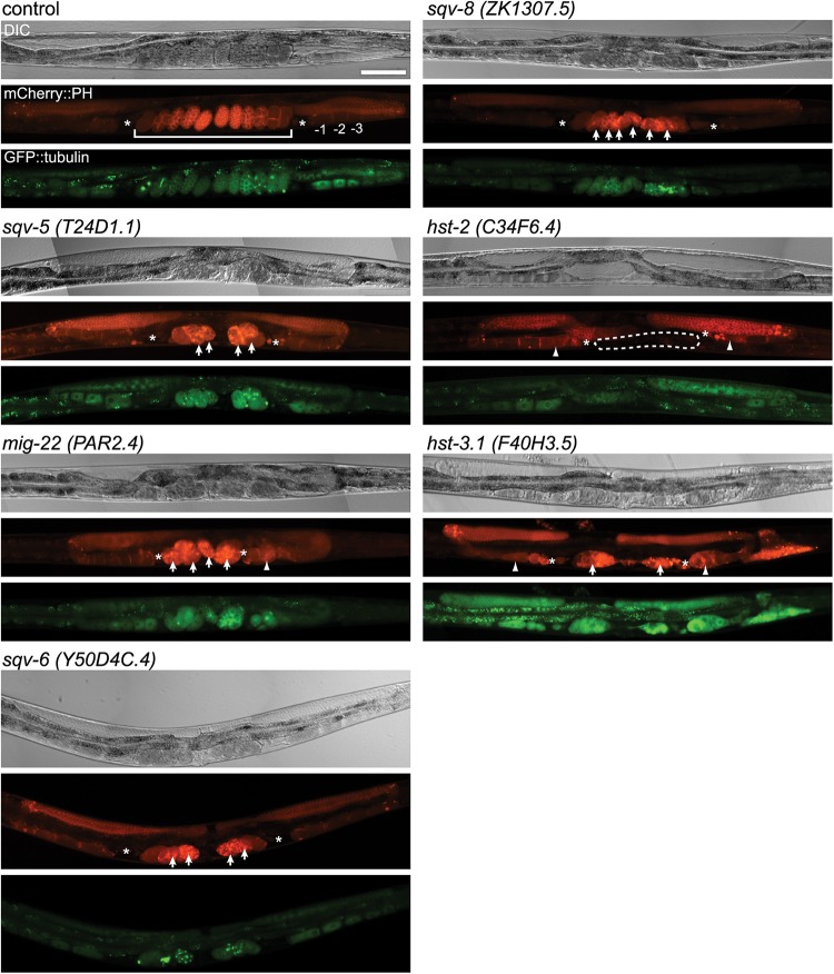
Fig. 1.
RNAi-mediated knockdown of several glycosyltransferases and sulfotransferases showed germline phenotypes. In each set of photographs, the upper panel shows the Nomarski DIC micrograph, the middle panel shows the PH domain detected by mCherry (red) and the bottom panel shows tubulin detected by gfp (green). In the mCherry::PH panel, the numbers indicate the normal oocytes, where −1 is the most proximal, and the brackets indicate the normal uterus (control); arrow: embryonic lethal (sqv-5, mig-22, sqv-6, sqv-8 and hst-3.1); arrowhead: oocyte morphology variant (mig-22, hst-2 and hst-3.1); dashed line: no eggs in uterus (hst-2), asterisks; spermatheca, scale bar: 100 μm.

