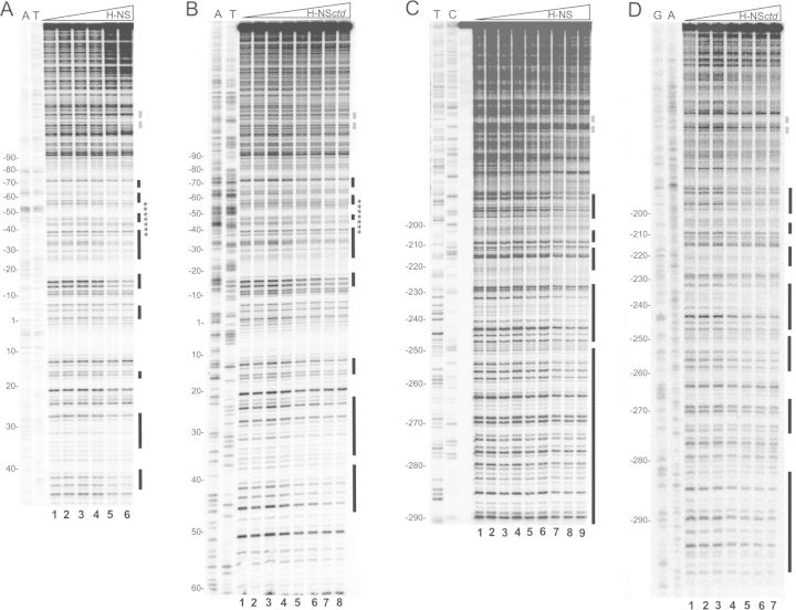
Figure 1.
DNase I footprinting of the virF promoter with H-NS and H-NSctd at equilibrium. A 450 bp DNA fragment containing virF promoter labeled on the non-coding (A and B) and coding (C and D) strands was incubated with DNase I and H-NS or H-NSctd as described in Materials and Methods. The concentration of the proteins was: (A) 0, 0, 23, 46, 114, 230 nM of H-NS dimers from lanes 1 to 6, respectively; (B) 0, 0, 13, 26, 52, 104, 157, 208 μM of H-NSctd monomers from lanes 1 to 8, respectively; (C) 0, 0, 23, 46, 69, 92, 114, 138, 230 nM of H-NS dimers from lanes 1 to 9, respectively; the lane preceding the samples subjected to enzymatic digestion contains the undigested DNA fragment. (D) 0, 13, 26, 52, 104, 157, 208 μM of H-NSctd monomers from lanes 1 to 7, respectively. T, C, A and G lanes represent sequencing reactions performed using primer GEN-348 for A and B and primer GEN-349 for B and D. Black bars indicate the protected sites; stars indicate the sequence of the 22mer fragment analyzed by NMR spectroscopy; broken gray lines indicate the center of the main curvature found in the virF promoter region (41). Numbering is given according to the +1 transcription start site.

