
Figure 5.
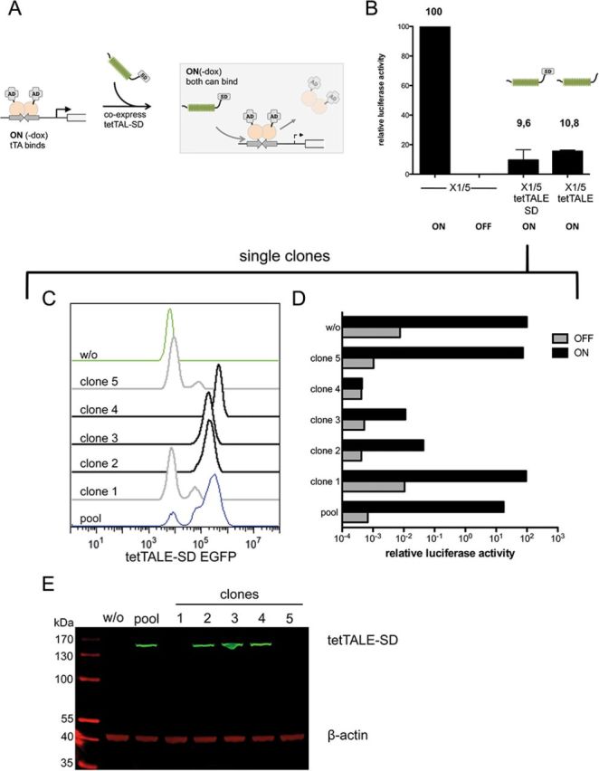
Competition between tTA and tetTALE/tetTALE-SD. (A) Experimental setup. X1/5 cells containing a Ptet-luciferase reporter and a tTA expression cassettes stably integrated were cultured under ON conditions (-dox) where tTA is bound to tetO. Cells were stably transfected with either tetTALE or tetTALE-SD expression construct containing a T2A-linked mCherry marker. (B) Analysis of luciferase activity of X1/5 cell pools (ON) transfected with either tetTALE or tetTALE-SD. Ptet-mediated luciferase activation of the TALE-negative parental cell lines with bound tet activator only was set to 100. Shown are mean values of three independent experiments with standard deviation. (C) Clones isolated from tetTALE-SD-transfected X1/5 cells were analyzed for tetTALE-SD linked GFP expression along with untransfected X1/5 cells (w/o) and the originating pool. (D) Single clones isolated from tetTALE-SD-transfected X1/5 cells grown under ON conditions were analyzed for luciferase activity 7 days after the switch of dox conditions from ON (-dox) to OFF (+dox). Ptet-mediated luciferase activation of the TALE-negative parental cell lines with bound tet activator only was set to 100. (E) Immunoblot analysis of single clones isolated from tetTALE-SD-transfected X1/5 cells grown under ON conditions to monitor tetTALE-SD expression levels. β-actin levels served as loading control.