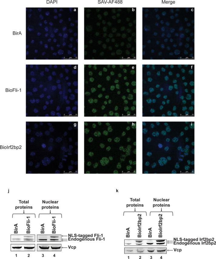
Figure 2.

Proper nuclear localization of the NLS-tagged proteins in MEL cells. (a–i) Immunofluorescence experiments in MEL/BirA (a, b, c), MEL/BioFli-1 (d, e, f) and MEL/BioIrf2bp2 (g, h, i) cells using either DAPI (a, d, g) or streptavidin conjugated with Alexa fluor 488 (b, e, h). The figure c, f and i show the merged picture. (j and k) Total (lanes 1 and 2) and nuclear (lanes 3 and 4) proteins were extracted from MEL/BirA (lanes 1 and 3), MEL/BioFli-1 (j, lanes 2 and 4) and MEL/BioIrf2bp2 (k, lanes 2 and 4) and subjected to Western blot analysis. Membranes were probed using an antibody against the endogenous protein (top panel) or against Vcp (bottom panel, loading control).
