Figure 2.

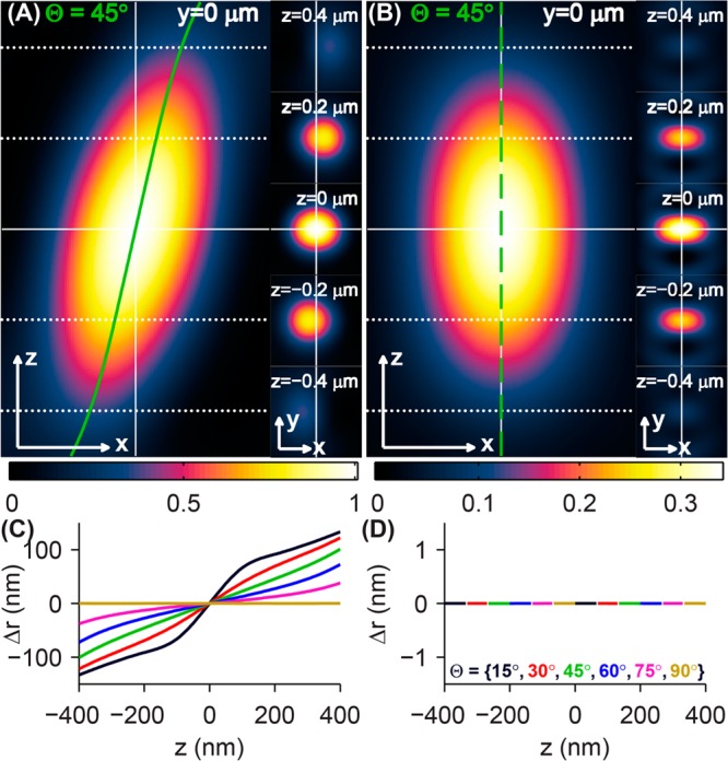
Simulated 3D PSFs and localization errors of rotationally fixed single dipole emitters. PSFs of a molecule oriented at {Θ = 45°,Φ = 0°} produced by (A) a microscope with a clear back focal plane and (B) a microscope with an azimuthal polarization filter. Cross sections of the 3D PSF in the xz (left) and xy (right) planes show the apparent shift of the dipole’s position as a function of defocus in the conventional microscope (solid green line in A), while the apparent lateral position of the molecule in the azimuthally polarized microscope remains fixed for all z positions (dashed green line in B). The intensity of the azimuthally polarized images is plotted relative to that of the clear-aperture microscope images. The apparent lateral position of an oriented molecule (black: Θ = 15°, red: Θ = 30°, green: Θ = 45°, blue: Θ = 60°, magenta: Θ = 75°, and gold: Θ = 90°) is computed as a function of defocus z for (C) the conventional microscope and (D) the azimuthally polarized microscope by fitting images to a 2D elliptical Gaussian function. The localization error Δr can be as large as ±100 nm for a defocus of 200 nm for conventional imaging. The imaging system with an azimuthal polarizer exhibits no localization error for any molecular orientation and any amount of objective lens defocus. Scale/axes arrows = 200 nm.
