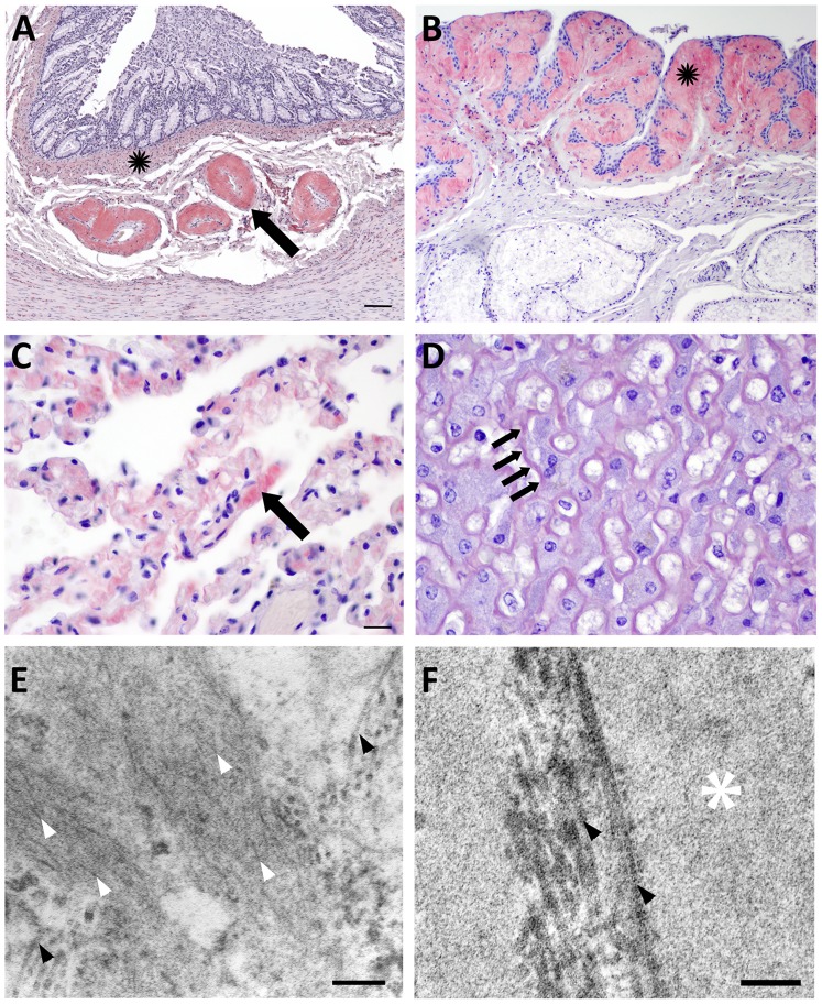
Figure 2. Congo red staining and electron microscopy of amyloid.
A: Amyloid markedly expands colonic submucosal arteries (arrow) and accumulates diffusely throughout the muscularis mucosae (black star). B: The epiglottis mucosa is undulating and hyperplastic and is expanded by submucosal amyloid (black star). C: Amyloid deposits thicken the capillary walls in the alveolar septae of the lung (arrow). D: Amyloid widens the Space of Disse in the liver (arrows) disrupting hepatic plates and isolating hepatocytes. E: The renal medullary interstitium from an amyloid positive fox shows long, thin, non-branching fibrils approximately 10 nm diameter (white arrowheads) arranged haphazardly amongst thicker bundles of collagen (black arrowheads). F: The renal medulla from an amyloid negative fox contains basement membrane (white star) and collagen (black arrowheads), but no fibrils. Scale bars = 200 µm (A, B); 50 µm (C, D); 200 nm (E); and 50 nm (F).

