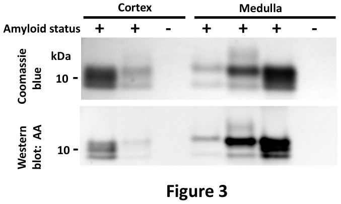
Figure 3. SDS-PAGE and western blot of the dominant insoluble protein in amyloid-laden kidneys.
Coomassie blue stain of insoluble proteins from the renal cortex and medulla show a dominant band at 12 kDa and one or two lower molecular weight bands at 10 and 8 kDa, consistent with full length and fragments of SAA. An immunoblot shows that 12 kDa and lower molecular weight bands react with anti-canine AA antibody.

