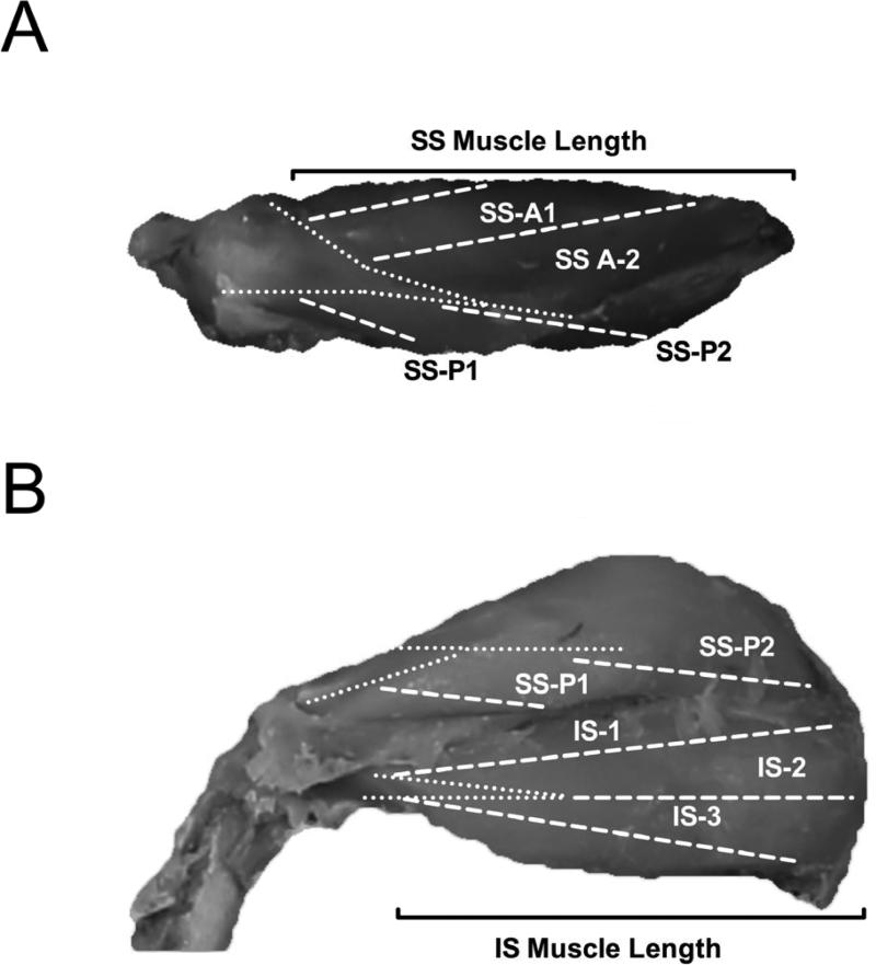
Figure 1.
Representative superior (A) and posterior (B) images of the rotator cuff. Dashed white lines represent the different sampling regions of the supraspinatus (SS) and infraspinatus (IS). Dotted white lines mark the borders of the deep tendons. Solid black brackets denote the muscle length for the supraspinatus (A) and infraspinatus (B). A=Anterior; P=Posterior.

