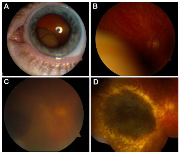
Fig. 1.
A 48-year-old woman had a large uveal melanoma with basal dimensions 12 mm × 11 mm, and height of 10.6 mm. Fine-needle aspiration biopsy of the tumor was performed at the time of Iodine-125 plaque radiotherapy, revealing a class 1 gene expression profile. Sixty-nine months post-treatment, the patient remains metastasis free. Baseline tumor findings seen on external (A) and fundus examination (B). C: One week following I-125 plaque radiotherapy, she developed pain and was found to have a significant uveitic response with vitritis. D: Two years after plaque radiotherapy, the tumor is completely flat with no residual uveitis

