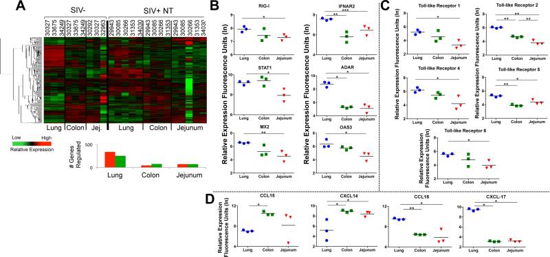
Figure 2. Distinct profiles of immune response associated gene expression in the lung and GI mucosa of therapy-naïve SIV infected animals and healthy controls.
(A) Hierarchical clustering of the genes with increased or decreased expression in the lung, colon, and jejunum of untreated SIV infected animals. (SIV+ NT indicates SIV+ animals without therapy). Baseline levels of transcription of genes associated with (B) type I IFN response (RIG-I, IFNAR2, STAT-1, ADAR, MX2, OAS3), (C) pathogen associated molecular patterns, or PAMPS (TLR1, 2, 4, 5, and 8); and (D) trafficking (CCL15, CXCL14, CCL18, CXCL17) are shown in the lung (blue circles), colon (green squares), and jejunum (red triangles) tissues of healthy macaques (n=3) were determined by microarray analysis. *p-value<0.05, **p-value<0.01, ***p-value<0.001

