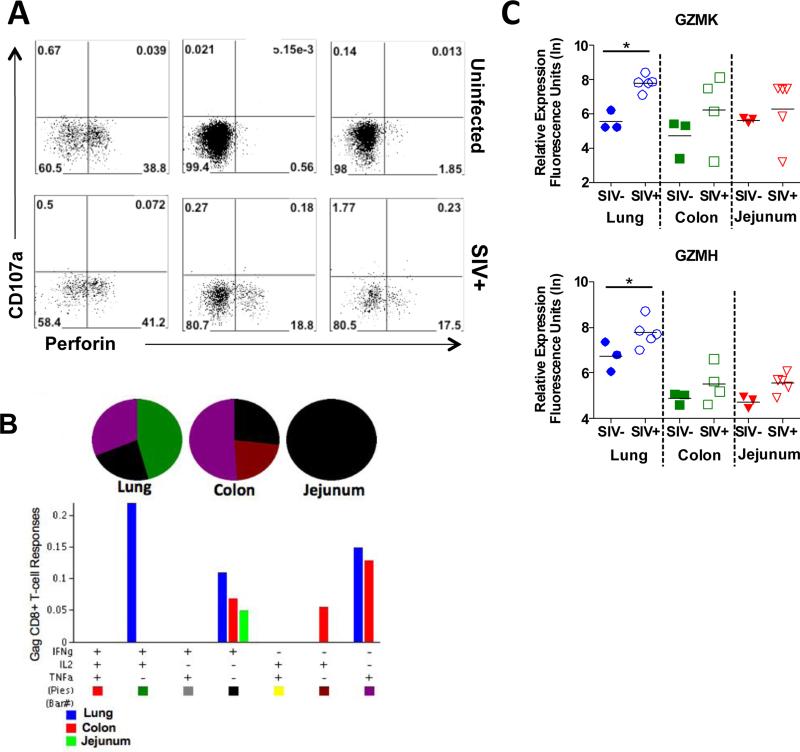
Figure 8. Comparison of lung and GI tract CD8+ T-cell responses in SIV infected macaques.
(A) Flow cytometric analysis of changes in CD8+ T-cell populations in the lung, colon, and jejunum expressing perforin and/or the degranulation marker, CD107a in response to SIV infection. A representative animal shown, (n=5). (B) In vitro production of IFNγ, IL-2, and TNFα in response to SIV gag stimulation was evaluated in CD8+ T-cells isolated from the lung, colon, and jejunum at 10 weeks post SIV infection (n=5). (C) Changes in the expression of granzymes K and H in the lung, colon, and jejunum during chronic stage SIV infection were determined by microarray analysis. *p-value<0.05

