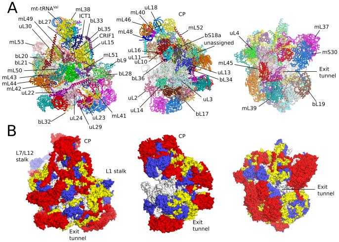
Fig. 1. Overview of human mt-LSU.
(A] Location of proteins in the human mt-LSU, showing (from left to right) solvent-facing, side and exit tunnel views. (B) Views as in A, proteins conserved with bacteria (blue), extensions of homologous proteins (yellow) and mitochondria-specific proteins (red). rRNA is shown in gray.

