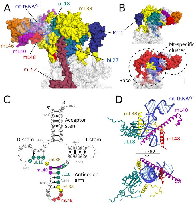
Fig. 3. The central protuberance containing mt-tRNAVal.
(A) Relative locations of proteins and mt-tRNAVal in the central protuberance. (B). View of A rotated by 180°, colored by proteins (top) and conservation (bottom) in accordance with Fig. 1. (C) Secondary structure of mt-tRNAVal. Modeled nucleotides are circled, and those interacting with surrounding proteins are colored. (D) The anticodon arm of mt-tRNAVal (blue) interacts extensively with proteins, whereas the acceptor arm is solvent exposed.

