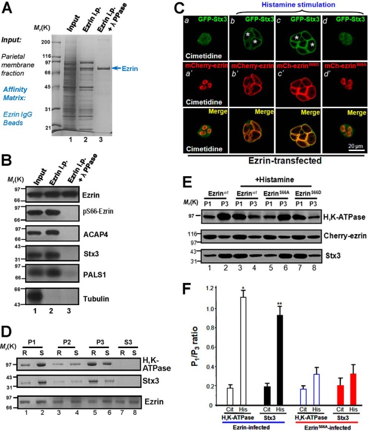
FIGURE 1.
Phosphorylation of ezrin at Ser-66 is essential for Stx3 and H,K-ATPase translocation to the apical plasma membrane. A and B, the plasma membrane-enriched P1 fraction was extracted with TX-100, and the extract was incubated with ezrin antibody-conjugated beads as described under “Materials and Methods.” The beads were washed successively with PBS before eluting ezrin with 0.2 m glycine, pH 2.3. All samples were separated by SDS gel electrophoresis. The proteins were either stained with Coomassie Blue (A) or transferred onto nitrocellulose membrane for Western blotting (B). Western blots were probed with an anti-ezrin antibody (top panel), anti-Ser(P)-66 antibody (second panel), anti-ACAP4 antibody (third panel), anti-Stx3 antibody (fourth panel), anti-PALS1 antibody (fifth panel), and anti-tubulin antibody (bottom panel). i.p., immunoprecipitate; λ PPase, λ-protein phosphatase. C, this montage represents confocal images collected from resting and secreting gastric parietal cells doubly stained for GFP-Stx3 (green), mCherry-ezrin (red), and their merged images. Ezrin is mainly located in the apical plasma membrane of parietal cells (seen as rings) in a pattern suggestive of the apical plasma membrane invaginations that form the intracellular canaliculi (b′). Labeling of Stx3 is mainly located in the cytoplasm of parietal cells as they associate with tubulovesicle (a′), whereas a lesser degree of co-localization is seen in apical membrane. However, stimulation induces remodeling of the apical membrane, seen as a dilation of apical vacuoles in the parietal cells (b′ and c′). Labeling of Stx3 is mainly located in the dilated apical vacuole membrane (c, asterisk), which is superimposed onto that of mCherry-ezrin distribution in the Merge. Note that histamine treatment maximally stimulates the dilation of apical vacuole membrane of mCherry-ezrinS66D-expressing cells but failed to dilate apical vacuole membrane of mCherry-ezrinS66A-expressing cells (d′). Bar, 15 μm. D, Western blotting analyses of ezrin, Stx3, and H,K-ATPase (α-subunit) of subcellular fractions derived from resting (R) and stimulated (S) gastric glands. Note that stimulation enriches the protein levels of H,K-ATPase and Stx3 in P1 fraction. E, Western blotting analyses of ezrin, Stx3, and H,K-ATPase (α-subunit) of subcellular fractions derived from resting and stimulated gastric glands infected with mCherry-ezrin adenovirus (wild type, ezrinS66A, and ezrinS66D). F, quantification of the α-subunit of H,K-ATPase and Stx3 proteins from P1 (plasma membrane-enriched) and P3 (tubulovesicle-enriched) fractions. The measurements were expressed as P1/P3 ratio. All data are given as the means ± S.E. (error bars) of four preparations. Cit, cimetidine; His, histamine.

