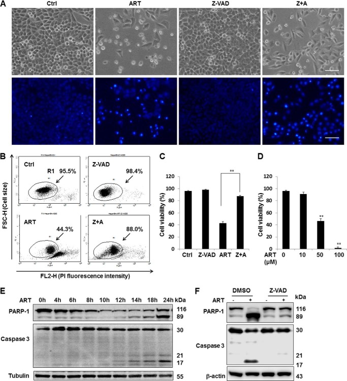
FIGURE 1.
ART induces apoptosis in cancer cells. A, HeLa cells were treated with ART (50 μm) with or without Z-VAD-fmk (20 μm) for 48 h. Morphological changes of ART-induced cell death were observed under a light microscope (top) or by using a fluorescent microscope after incubation with Hoechst dye for 30 min (bottom). Scale bar, 200 μm. B, HeLa cells were treated as indicated in A, and the cell death was quantified by flow cytometry using PI (5 μg/ml) staining (R1 area, representative of viable cells). C, statistical analysis of the percentage of viable cells of three independent experiments performed as in B (mean ± S.D. (error bars)) (**, p < 0.01, Student's t test). D, HeLa cells were treated with the indicated dose of ART, and cell death was quantified by flow cytometry after PI staining as described earlier (**, p < 0.01, Student's t test). E, HeLa cells were collected after treatment with ART (50 μm) for the indicated period of time, and Western blotting was then performed for the detection of various indicated markers of apoptosis. β-Actin and tubulin served as loading controls. F, HeLa cells were treated with ART (50 μm) alone or combined with Z-VAD-fmk (20 μm) together for 24 h followed by Western blotting to detect the apoptosis markers.

