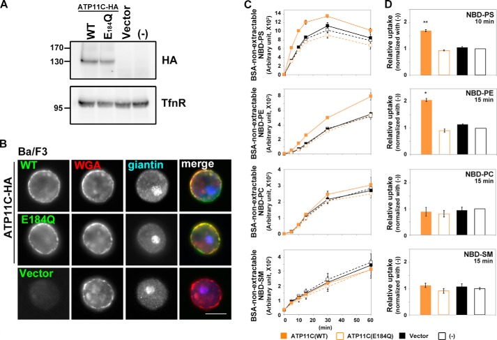
FIGURE 1.
Flippase activities of ATP11C across the leaflets of the plasma membrane in Ba/F3 cells. Parental Ba/F3 cells (−) or Ba/F3 cells infected with empty retrovirus vector (Vector) or recombinant virus vector encoding HA-tagged ATP11C(WT) or ATP11C(E184Q) were either processed for immunoblot analysis with antibodies against HA and TfnR (as an internal control) (A) or stained with Alexa Fluor 555-conjugated wheat germ agglutinin (WGA) to visualize the plasma membrane followed by immunostaining for HA and giantin (a Golgi marker) (B). Scale bar, 10 μm. C, Ba/F3 cells were incubated with the indicated NBD-lipids at 15 °C for the indicated times (x axis). After extraction with fatty acid-free BSA, the residual fluorescence intensity associated with the cells was determined by flow cytometry. Graphs are representative of two independent experiments, and results display averages from triplicates ±S.D. D, -fold increase of NBD-lipid uptake compared with parental cells (−) is shown at the 10- (NBD-PS) or 15-min (NBD-PE, -PC, and -SM) time point from C. Graphs are representatives of three independent experiments, and results display averages from triplicates ±S.D. (*, p < 0.0001; **, p < 0.0005). Error bars represent S.D.

