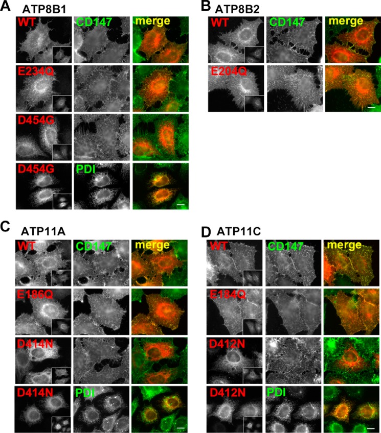
FIGURE 2.
Plasma membrane localization of P4-ATPases and their ATPase-deficient mutants in HeLa cells. HeLa cells were transiently co-transfected with expression vectors for FLAG-tagged CDC50A and HA-tagged ATP8B1(WT), ATP8B1(E234Q), or ATP8B1(D454G) (a BRIC mutant, see Fig. 7) (A); ATP8B2(WT) or ATP8B2(E204Q) (B); ATP11A(WT), ATP11A(E186Q), or ATP11A(D414N) (C); or ATP11C(WT), ATP11C(E184Q), or ATP11C(D412N) (D). Before fixation, cells were incubated with Alexa Fluor 488-conjugated anti-CD147 antibody for 5 min at room temperature to label the plasma membrane. The fixed cells were then incubated with anti-HA and anti-FLAG (M2) antibodies followed by Cy3-conjugated anti-rat and DyLight649-conjugated anti-mouse secondary antibodies. For protein-disulfide isomerase (PDI; an endoplasmic reticulum marker) staining, fixed cells were incubated with anti-HA, anti-FLAG, and anti-protein-disulfide isomerase antibodies followed by Cy3-conjugated anti-rat, Alexa Fluor 647-conjugated anti-rabbit, and Alexa Fluor 488-conjugated anti-mouse secondary antibodies. Insets indicate FLAG-CDC50A-expressing cells. Scale bars, 10 μm.

