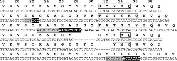
FIGURE 1.

Examples of the indels observed for a single antibody HC. Examples of the indels observed for a single antibody HC obtained when the antibody was expressed in vitro with AID in the absence of selection for improved antigen binding are shown. The starting amino acid and DNA HC sequence (top) is shown together with five in-frame indels and one indel producing a frameshift (bottom). CDR H1 regions are highlighted with a box; deletions are indicated by dashes, and Kabat numbering is shown across the top. For insertions, the upstream sequence is shown in underlined light gray type, and the resulting duplicated sequence is highlighted in black.
