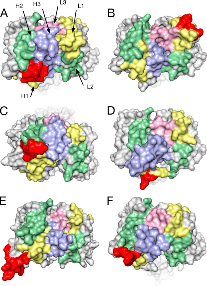
FIGURE 6.
Comparison of human antibody structures containing insertions. Molecular surface representation of the crystal structures of Fabs of human antibodies with different specificities featuring insertions ranging from 4 to 9 amino acids. CDRH1 (yellow), CDRH2 (green), CDRH3 (blue), CDRL1 (yellow), CDRL2 (green), CDRL3 (pink), and insertions (red) are shown in the same orientations in a top view onto the combining site. A, APE1551, an anti-human βNGF antibody with a 9-residue insert in CDRH1 (PDB code 4NWU). B, bH1, a dual specific antibody to HER2 and VEGF, 4-residue insert in CDRL1 (PDB code 3BDY) (47). C, PGT127, an anti-gp120 antibody that recognizes conserved glycan residues, 6-residue insert in CDRH1, 4-residue deletion in CDRL1 (PDB code 3TWC) (15). D, C05, an anti-influenza A hemagglutinin antibody, 5-residue insert in CDRH1 (PDB code 4FP8) (17). E, VRC06, an anti-gp120 antibody, 7-residue insert in heavy chain FR3, 1-residue deletion in CDRL1 (PDB code 4JB9) (48). F, PGT135, an anti-gp120 antibody, 5-residue insert in CDRH1 (PDB code 4JM2) (49).

