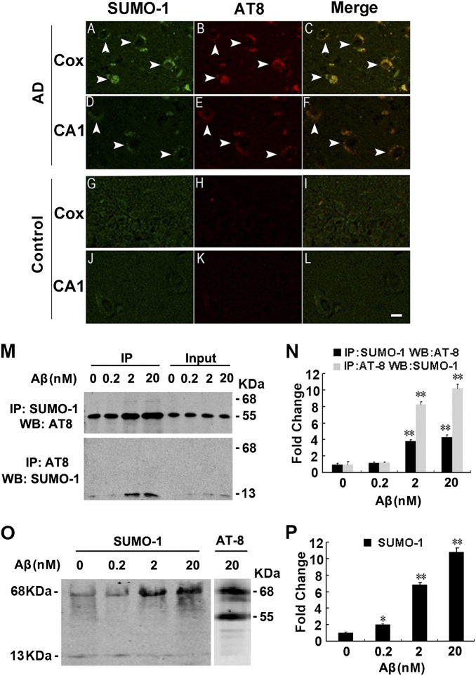
Fig. 5.
Colocalization of SUMO-1 with the phosphorylated tau in the AD brains. (A–L) Double-staining of SUMO-1 and the phosphorylated tau (AT8) in the cerebral cortex (Cox) and hippocampal CA1 of the AD brains (A–F) and the age-matched controls (G–L). (Scale bar, 20 μm.) (M and N) Rat primary hippocampal neurons (14 DIV) were treated with 0, 0.2, 2, 20 nM of Aβ1–40 for 24 h. Co-IP)/Western blotting (WB) (M) and quantitative analyses (N) were performed by using AT8 and SUMO-1 antibodies (note that sample boiling required in the co-IP procedure causes dropout SUMO, therefore, only ∼13-kDa band of endogenous SUMO-1 was observed in M). (O and P) Rat primary hippocampal neurons (14 DIV) were treated as described above, then the samples were extracted with RIPA buffer without boiling for Western blotting. After separated by SDS/PAGE on the same gel, the membrane was cut into two and immunostained respectively with SUMO-1 and AT8 (note that the 68-kDa band stained by both SOMU-1 and AT8 represents the endogenously SUMOylated tau induced by Aβ exposure). *P < 0.05, **P < 0.01 vs. DMSO (0 nM Aβ).

