Fig. 1.
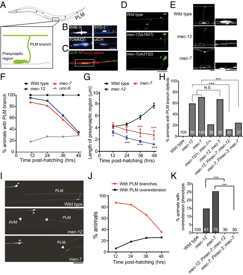
mec-12 and mec-7 are essential for the PLM branch stability. (A) Schematic diagram of C. elegans touch neurons. Anterior is to the left. (B) The PLM presynaptic varicosities were enriched in synaptic vesicles, the active zone protein SYD-2, the mitochondria, and the synapse-specific F-actin. The PLM neurite was labeled by zdIs5(Pmec-4::GFP) or jsIs973(Pmec-7::mRFP) and pseudocolored in blue and synaptic markers in white. (Scale bar, 10 μm.) (C) Confocal image showing that the PLM presynaptic varicosities (red) opposed GLR-1 clusters (green) of the postsynaptic glutamatergic interneurons. (Scale bar, 5 μm.) (D) Confocal images of the PLM branch (arrow) in L4 larvae. Asterisks, PVM neurons; arrowheads, PLM processes. (Scale bar, 20 μm.) (E) The PLM presynaptic varicosities, visualized with zdIs5(Pmec-4::GFP), at the L4 stage. Boxed regions were highlighted. (Scale bar, 10 μm.) (F) Quantification of animals with intact PLM branches at different larval stages. (G) The length of the PLM presynaptic varicosities at different larval stages. Error bar, SEM **P < 0.01; ***P < 0.001, Mann–Whitney U test. (H) Quantification of L4 animals with PLM branch defects. tm5083 and e1507 were other null alleles for mec-12 and mec-7, respectively. ***P < 0.001, two-proportion z test. N.S., not significant. (I) Confocal images of PLM neurite overextension in L4 larvae. Arrowheads, ventral migration of the PLM process tip to reach the ventral nerve cord; asterisks, ALM soma. (J) Quantification of PLM branch retraction and neurite overextension in mec-7 animals at different larval stages. n > 30. (K) Quantification of L4 animals with PLM neurite overextension. ***P < 0.001, two-proportion z test.

