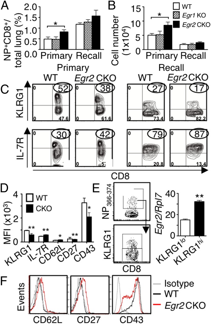
Fig. 6.
A higher percentage of antigen-specific CD8+ T cells have a memory phenotype in Egr2 CKO mice than in WT mice after influenza infection. (A and B) Lung cells were stained with NP366–374 pentamer. Frequency (A) and absolute NP+CD8+ cell numbers (B) at peak primary and recall infection time points. (C) Flow cytometric profile of KLRG1hi and IL-7Rhi antigen-specific CD8+ T cells in primary (Left) or recall (Right) infection models. (D) Mean fluorescent intensity (MFI) of KLRG1, IL-7R, CD62L, CD27, and CD43 from antigen-specific CD8+ T cells after primary infection. (E) Antigen-specific KLRG1lo and KLRG1hi CD8+ T cells were sorted and Egr2 RNA expression on the two populations. (F) FACS profile of CD62L, CD27, and CD43 expression in antigen-specific CD8+ T cells. Data are for 24 mice per group, combined from four experiments (mean ± SD) in A, B, and D or from representative experiments in C, E, and F.

