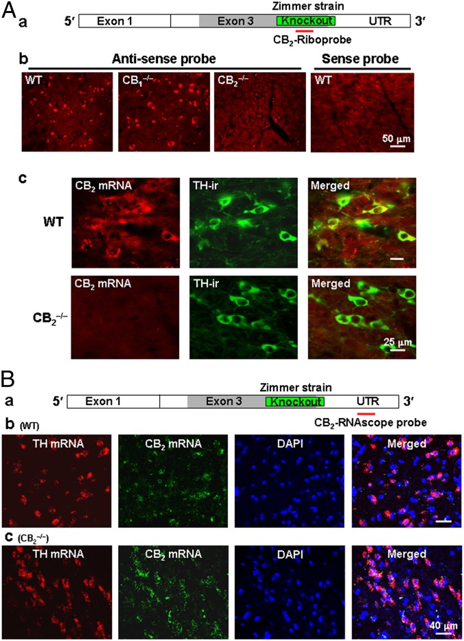
Fig. 2.
CB2 mRNA expression in VTA DA neurons by ISH assays. (A, a) CB2A mRNA and the location detected by a CB2-riboprobe. (A, b) The antisense, but not the sense (control), riboprobe detected CB2 mRNA in midbrain neurons in WT and CB1−/− mice, but not in CB2−/− mice. (A, c) Double-label fluorescent images of CB2 mRNA (by ISH) and TH (by IHC) staining, illustrating CB2 mRNA staining in individual TH-positive VTA DA neurons in WT mice, but not in CB2−/− mice (Zimmer strain). (B, a) CB2A mRNA and the location detected by a CB2-RNAscope probe. (B, b and c) CB2-RNAscope probe-detected CB2 mRNA in VTA DA neurons in WT and CB2−/− mice. This probe detected CB2 mRNA in CB2−/− mice because it targets the downstream UTR region rather than the upstream gene-deleted region. Scales are shown in the figures. Also see Fig. S1.

