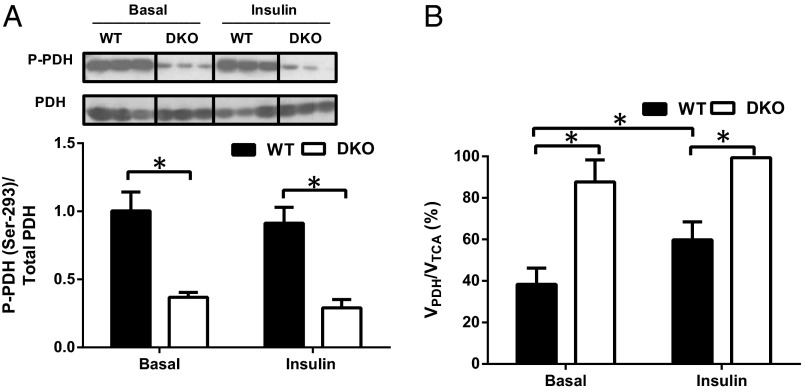
Fig. 1.
Decreased phosphorylation of the PDH complex subunit E1α increases PDH flux in the skeletal muscle of DKO mice. (A) Representative immunoblots of the Ser293-phosphorylated form of the PDH complex E1α from WT and PDK2/PDK4 DKO mice. Histograms constructed from the data obtained from Western blot analysis. (B) Relative PDH flux (VPDH/VTCA) in the skeletal muscle of WT and DKO mice after overnight fasting (basal) and at the end of the hyperinsulinemic-euglycemic clamp (insulin). Data are mean ± SEM. n = 8 mice per group.

