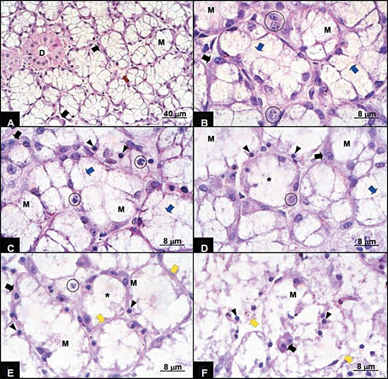
FIGURE 1.
Sublingual gland A - 0 h: the glandular structure presents integrity, exhibiting mixed acini with large mucous cells (M) and small half-moon serous cells (full arrow) and intralobular ducts (D) interlaced by stroma (reed arrow). HE; B - 3 and 6 h: mixed acini exhibiting some mucous cells (M) presenting loss of integrity of their external limit (blue arrow) and integral serous cells (full arrow); C - 12 h: mixed acini exhibiting mucous cells (M) presenting loss of integrity of their external limit (blue arrow), karyorrhexis (circle) and pyknosis (arrowhead) and serous cells presenting karyorrhexis (full arrow). HE; D - 12 h: mixed acini exhibiting mucous cells (M) presenting loss of integrity of their external limit (blue arrow) and others showing total disintegration of the external limit (asterisk), karyorrhexis (circle) and pyknosis (arrowhead) and serous cells presenting pyknotic nuclei (full arrow). HE; E - 24 h: Disorganization of glandular in relation to the previous figure, exhibiting mixed acini retaining their limits (yellow arrow), exhibiting mucous cells (M) presenting total and disintegration of the external limit (asterisk), karyorrhexis (circle) and pyknosis (arrowhead) and serous cells presenting pyknotic nuclei (full arrow). An acinus showing a single remaining nucleus was noted. HE; F - 24 h: Complete disorganization of the glandular structure, exhibiting acini presenting external limit rupture (yellow arrow) complete disorganization of the cellular content and integral serous cells dispersed in the amorphous resulting from mucous cell destruction. Hematoxylin-eosin

