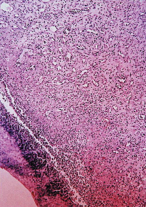
Figure 3.

Microscopic aspect showing an oral mucosa consisting of continuous parakeratinized, stratified pavement epithelium covered with a serofibrinous membrane. The underlying granulation tissue was rich in blood vessels, fibroblasts and macrophages. Skeletal striated muscle fibers were observed in the deep layers (Original magnification x 25)
