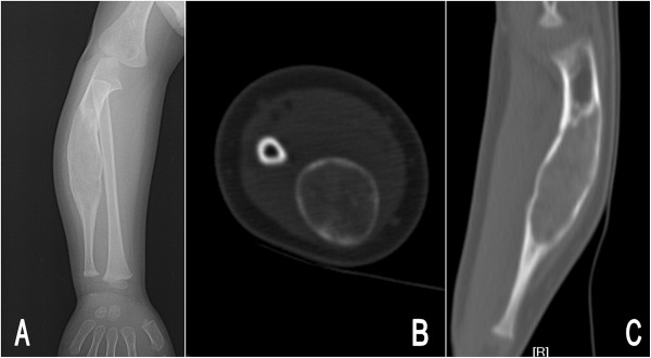
Figure 1.

Preoperative images of the right forearm in patient 1. Posteroanterior radiograph (A) showed a fusiform-shaped, expansile ground-glass lesion in the ulna. Axial plane (B) and coronal reconstructed computed tomography (C) revealed the cortex was thinning and the area of lesion was homogeneous, with an average computed tomography value of 123 Hu.
