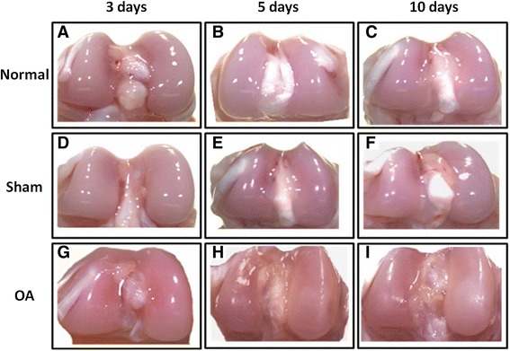
Figure 1.

Macroscopic changes in the articular cartilage of rat during the early stages of OA. Femoral condyles from normal, sham, and early induced OA as viewed with a stereomicroscope. (A-C) Femoral condyles from normal, (D-F) sham, and (G-I) OA-induced (OA 3, 5, and 10 days) rats.
