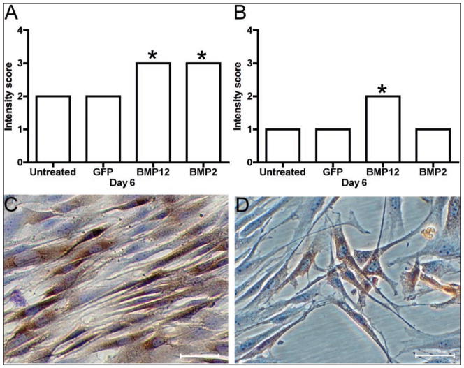
Figure 6.
Type I collagen in equine SDFTNs (A, C) and BMDMSCs (B, D). On cell culture day 6, SDFTN-BMP12 and BMP2 cells had significantly (P < 0.001) greater type I collagen staining intensity, compared with that of the other groups (A). On cell culture day 6, BMDMSC-BMP12 cells had significantly (P < 0.001) greater staining intensity, compared with that of the other groups (B). C—Photomicrograph of SFTN-BMP12 cells containing type I collagen. Immunohistochemical stain; bar = 20 μm. D—Photomicrograph of BMD-MSC-BMP12 cells containing type I collagen. Immunohistochemical stain; bar = 20 μm.

