
Figure 1.
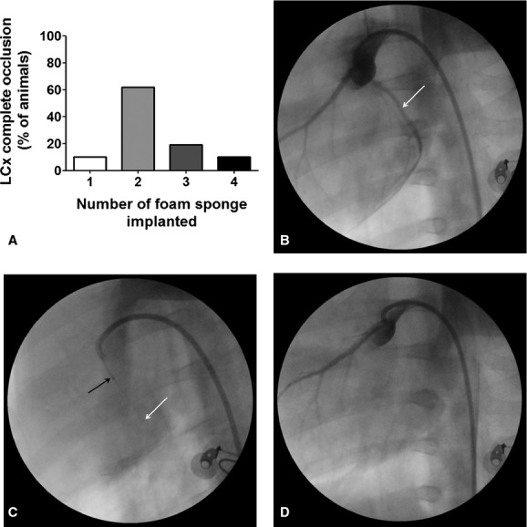
Foam sponge implanted in LCx. (A) Number of pieces of foam sponge to completely occluded the lumen of LCx. (B) First, a basal angiography was performed to localize the coronary tree. (C) after localized the target (proximal portion) a 0.014 guidewire (white arrow) was carried up to distal portion of LCx. Through the guidewire and assisted by a balloon catheter (black arrow) pieces of foam sponge were placed into LCx lumen. (D) Five to ten minutes after occlusion catheters and guidewire were removed from artery.