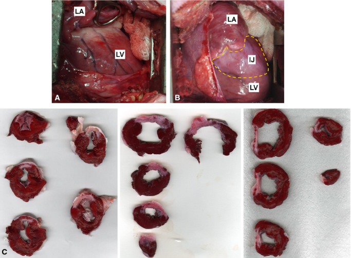
Figure 4.

Anatomopathological assessment of LV 30 days after LCx occlusion. (A) Open‐chest view of LV posterior wall before LCx. (B) Open‐chest view of LV posterior wall 30 days after LCx occlusion. Note injured area in white (discontinuous yellow points). (C) Sliced sections of LV from three different pigs. Note that the injured areas are similar. LV, left ventricle; LA, left atrium; IJ, injured area.
