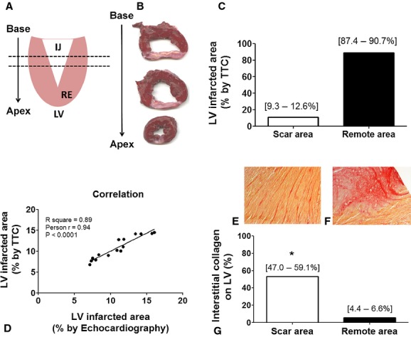
Figure 5.

Anatomopathological follow‐up 30 days after LCx occlusion – histological confirmation and quantification of infarcted area. (A) After euthanasia the heath was isolated, LV dissected and sliced base to apex as schematic figure. (B) The injury extended from the base to the apex forming an inverted triangle clearly observed in LV slices stained with TTC. (C) LV injured perceptual area was homogeneous for all the animals (n = 15) and (D) showed strong correlation with echocardiographic measures (n = 15). Furthermore, a significant increase in interstitial collagen in (E) remote and (F) injured areas were (G) quantified versus the amount of collagen in healthy animals LV (2% of interstitial collagen all over the LV; P < 0.001). IJ, injured area; RE, remote area.
