Figure 4.
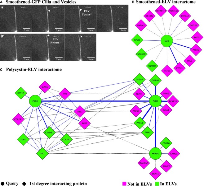
Fluorescently labeled ELVs appear to interact with renal primary cilia. (A) Still image from a spinning disk confocal movie (Supplemental movie 1) of Smoothened‐YFP MDCK cells showing small Smoothened‐containing vesicles (arrowheads) in the extracellular region interacting with renal primary cilia. A'. Small vesicles appear to interact with primary cilia. B'. A small vesicle appears to be released to the extracellular environment from the tip of the primary cilium. (B) First‐degree interaction map for Smoothened illustrating which proteins are present (green) and absent (magenta) in human ELVs. (C) First‐degree interaction map for PKD1 which encodes Polycystin‐1, PKD2 which encodes Polycystin‐2, and PKDH1 which encodes Fibrocystin, illustrating which proteins are present (green) and absent (magenta) in human ELVs. The solid blue line refers to a physical interaction that has been reported, and the dotted line refers to a genetic interaction.

