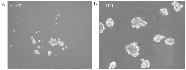
Figure 2.

Phase contrast microscopy, demonstrating (A) CD133+ cells gradually formed sphere colonies of different sizes and irregular shapes following approximately one week of culturing and (B) CD133+ cells were exclusively formed of cellular clusters of tumor spheres following approximately one month of culturing in serum-free medium containing various growth factors. CD, cluster of differentiation.
