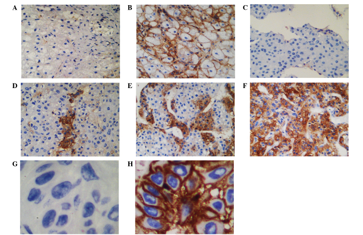
Figure 1.
Immunohistochemical staining of human leukocyte antigen-F (HLA-F) expression in hepatocellular carcinoma (HCC) lesions and normal liver tissues. Immunohistochemically (A) negative and (B) positive expression of HLA-F in normal liver tissues. (C) Negative, (D and E) ≤50% positive and (F) >50% positive HLA-F expression in HCC lesions. HLA-F expression was considered as negative when the percentage of stained cells was ≤5%. Original magnification, ×100. (G) Negative and (H) positive skin cancer lesion controls. Original magnification, ×400.

