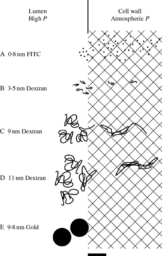
Fig. 10.

Diagrammatic representation of molecular movement into the primary wall at P of 0·5 MPa found in living Chara cells. (A) Molecules smaller than wall interstices (cross-hatched). (B) Polysaccharides near the dimensions of the interstices. (C) Polysaccharides having diameter as much as twice the dimensions of the interstices, starting an end into the wall aided by P. (D) Polysaccharides having diameter more than twice the dimensions of the interstices and having whole molecule or segment of molecule being deformed by P. (E) Gold colloids at about twice the dimensions of the interstices cannot be deformed and do not enter wall significantly. The molecules, colloids and wall interstices are drawn to scale. Scale bar = 10 nm.
