Fig. 3.
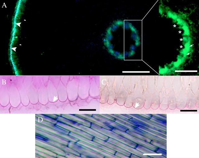
(A) Cross-section at 5 mm from the root tip shows autofluorescence of outer tangential epidermal cell walls (arrowheads). Protoplasts of endodermis (arrows) can be recognized on the outer edge of autofluorescent area external to vascular tissues (asterisks); not stained; fresh section. Scale bar = 50 μm. (B) Lipidic material was detected in the outer epidermal cell walls (arrow); fresh section (5 mm from root tip) stained with sudan red 7B. Scale bar = 25 μm. (C) Lignification of outer epidermal cell walls (arrow); fresh section; HCl–phloroglucinol; 5 mm from tip. Scale bar = 25 μm. (D) Surface view of epidermis; fresh longitudinal section stained with toluidine blue, about 30 mm from tip. Scale bar = 50 μm.

