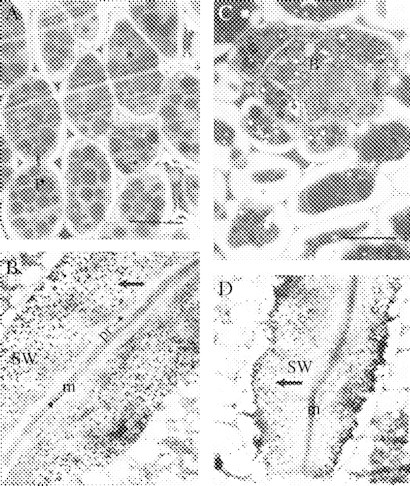
Fig. 5.

(A, B) Day 0 (16-h imbibed seeds). (A) Light microscopy of the storage mesophyll tissue of imbibed lupin cotyledons. Staining with methylene blue. Scale bar = 50 μm. (B) Ultrastructural view of a junction between two adjacent mesophyll cells from lupin cotyledons (×11 600). (C, D) Day 2. (C) Light microscopy of the storage mesophyll tissue, showing vascular bundle. Scale bar = 40 μm. (D) Ultrastructural view of a junction between two adjacent storage mesophyll cells (×11 600). Arrows in (B) and (D) point to gold particles. Labelling with the heat-treated, gold-complexed enzyme followed by staining with 2% uranyl acetate and 4% lead citrate. m, middle lamella; P, protein body; pr, primary wall; SW, storage wall; B, vascular bundle.
