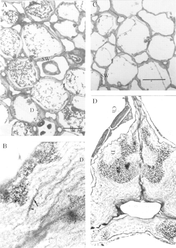
Fig. 7.

(A, B) Day 8. The dark regions in the wall are stained with galactanase–gold particles whereas the clear regions are not. (A) Storage mesophyll tissue during degradation of storage cell wall. (B) Ultrastructure of the storage cell wall during mobilization. The arrow points to fibrous material (possibly clusters of rhamnogalacturonan core molecules), which appears after degradation of galactan in vivo (×15 400). (C, D) Day 10. (C) Storage mesophyll tissue. Scale bar = 40 μm. (D) Ultrastructural view of a junction between three adjacent mesophyll cells (×4600). Labelling with the heat-deactivated gold-complexed enzyme followed by staining with 2% uranyl acetate and 4% lead citrate. Ch, chloroplasts; D, digestion pocket; SW, storage wall.
