
Fig. 1.
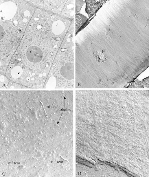
(A) Ultrathin section of recently divided cells just below the meristem of a Zea mays root tip. Note the recently synthesized transverse walls (thinner). The elongation axis will be perpendicular to this direction. (Unpublished micrograph, courtesy of Susette Mueller and R. Malcolm Brown, Jr.) (B) Freeze fracture showing the E fracture face (EF) of a large area of an elongating cell in the root of Zea mays. The direction of microfibril impressions and, hence, the direction of the orientation of the microfibrils themselves, is perpendicular to the axis of elongation. Note also a prominent pit field (pf) in the centre of the micrograph. Microfibril synthesis around this pit field gives clues that suggest a membrane flow mechanism in the plane of the fluid membrane may underlie and direct cellulose microfibril synthesis (see Mueller and Brown, 1982a, b). Evidence to support this hypothesis is based on the direction of microfibrillar tears through the plasma membrane where the terminal globules and direction of synthesis is revealed (see C). In addition, parallel cortical microtubules provide the general ‘channels’ for the membrane flow. Actin microfilaments are found perpendicular to the cortical microtubules and may be the source of motion to propel the directional motions of the fluid membrane. (Unpublished micrograph, courtesy of Susette Mueller and R. Malcolm Brown, Jr.) (C) E fracture face of the plasma membrane of an actively elongating cell in the root of Zea mays showing three prominent tears of microfibrils back through the outer leaflet of the plasma membrane (mf tear). Note that the ‘rip’ terminates at a hole where the microfibril is associated with the rosette TC. In this fracture face, only the globular regions of the tips are shown associated with the TCs (globules). Many other TCs which have not been torn through the plasma membrane are revealed, some in clusters. (Unpublished micrograph, courtesy of Susette Mueller and R. Malcolm Brown, Jr.) (D) Freeze fracture through the innermost layer of a growth wall from an elongating cell in the root of Zea mays. Note the change in pitch of the transverse walls, suggesting that during elongation, the general pitch of the direction of microfibril synthesis is gradually changing from transverse to longitudinal. (Unpublished micrograph, courtesy of Susette Mueller and R. Malcolm Brown, Jr.)