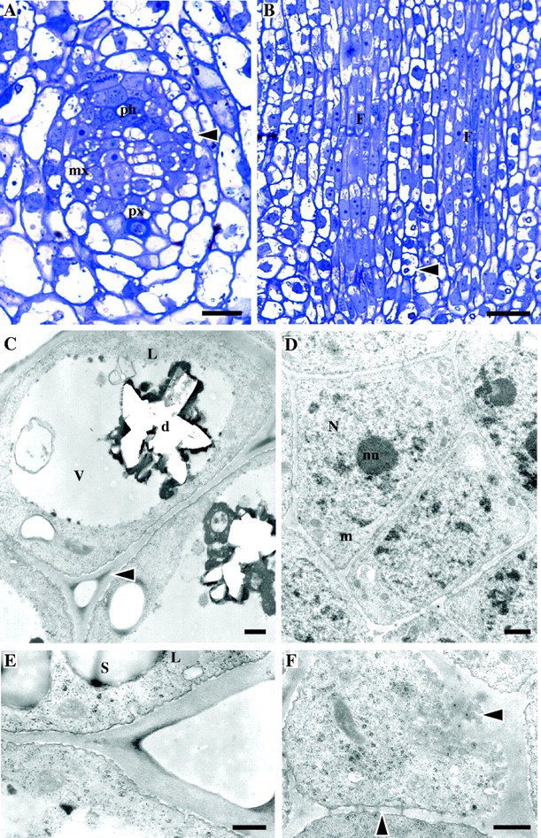
Fig. 1.

Stage 1 of development (un-elongated internodes close to apex of the immature shoot). Light (A, B) and TEM (C–F) micrographs. (A) TS of a provascular strand of an internode close to the apex showing differentiation of vascular elements. Ph, protophloem; px, protoxylem; mx, metaxylem; arrow, dividing fibre cells. (B) LS of a similar area showing the procambium strands which give rise to fibre cells (labelled ‘F’). The young fibres have a central protoplast and vacuolated ends. Note crystals in parenchyma cells (arrow). (C) Parenchyma cells (TS) with peripheral cytoplasm and large central vacuole (v) containing a hollow structure representing a druse (d). Intercellular spaces are present at cell corners, which are more electron-dense than the rest of the wall (arrow). Lipid globules (L) can be seen in cytoplasm. (D) Meristematic cells of the procambium (TS), which give rise to fibres, showing large nuclei (N) with prominent nucleoli (nu) and dense cytoplasm. Cells are surrounded by a continuous wall. The plasmalemma has an undulating appearance. (E) Detail of parenchyma cell wall showing vesicles fusing with the plasma membrane (TS). Starch grains (s) are present. L = lipid globule. (F) Glancing section of the end wall of a parenchyma cell where plasmodesmata can be seen in cross-section in the plasma membrane and in LS in lateral cell walls (arrows). Scale bars: (A) 50 μm, (B) 25 μm, (C–F) 0·5 μm.
