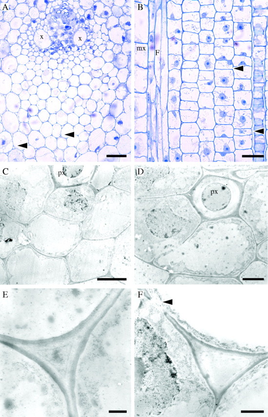
Fig. 2.

Stage 2 of development (internode 4 of the immature shoot). Light (A, B) and TEM (C–F) micrographs. (A) TS of early vascular bundle development. Some cells adjacent to xylem vessels (x) have prominent protoplasts. Intercellular spaces separate parenchyma cells in ground tissue and fibre cells, especially those in the main portion of the protoxylem fibre sheath (arrows). Parenchyma cells have a round or oval shape whereas fibres tend to be polygonal. (B) LS of a similar area showing elongating fibre cells (F) next to a differentiating metaxylem vessel (mx). Fibres have tapering tips, which grow intrusively at both ends of the cell. Most parenchyma cells continue to divide by periclinal divisions. Elongation of some parenchyma cells gives rise to long and short cells (arrows). (C) Developing fibre cells (TS) at the protoxylem (px) pole. Cells adjacent to the protoxylem element have large nuclei. (D) Detail of fibre cells (TS). (E) Cell wall of fibre cells from the central region of the protoxylem fibre sheath showing a newly deposited electron-dense layer on the cytoplasmic side. An intercellular space is developing, but it appears filled with unidentified material. (F) Cell wall between parenchyma cells showing similar characteristics to those of fibres. A primary pit field is present (arrow). Scale bars: (A, B) 50 μm, (C, D) 5·0 μm, (E, F) 1·0 μm.
